Compact Bone Diagram Microscope - 2 Microscopic Structure Of Compact Bone Download Scientific Diagram / The outer walls of the diaphysis cortex cortical bone are composed of dense and hard compact bone a form of osseous tissue.
byAdmin•
0
Compact Bone Diagram Microscope - 2 Microscopic Structure Of Compact Bone Download Scientific Diagram / The outer walls of the diaphysis cortex cortical bone are composed of dense and hard compact bone a form of osseous tissue.. A diagram of the anatomy of a bone, showing the compact bone. A cross section of decalcified compact bone is examined under brightfield illumination with the intel qx3 microscope. Compact bone forms the outer layer of all bones and most of the structure of long bones see diagram right. The ground substance of bone is arranged in concentrated layers (lamellae) round the small canals which run parallel to the long axis (shaft) of the bone. Proper use of a microscope.
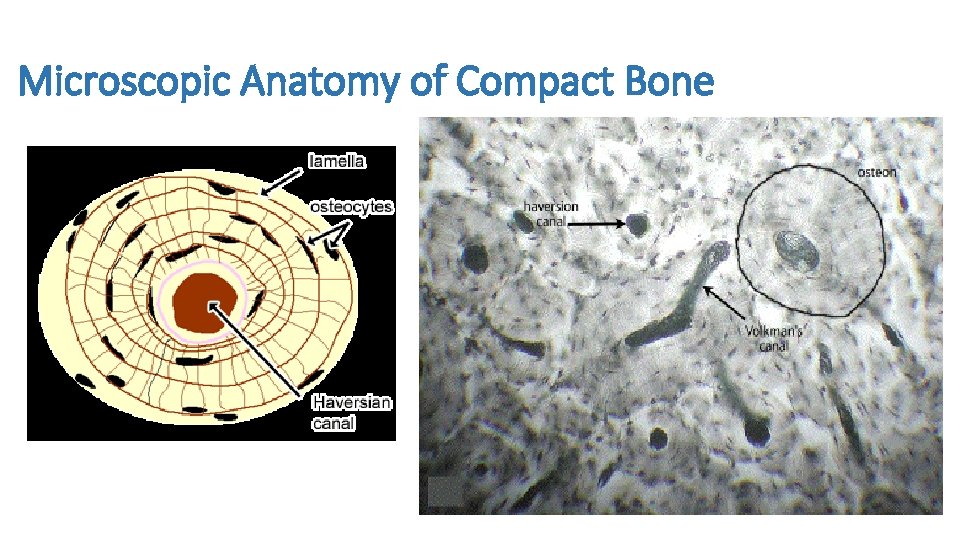
Each group of concentric circles (each tree) makes up the microscopic structural unit of compact bone called an osteon (this is also called a haversian. If you look at compact bone under the microscope, you will observe a highly organized arrangement of concentric circles that look like tree trunks. I love how this diagram includes the volkmann's canal, the haversian canal, the canaliculi and the priosteum. Zoo lab practical at high point university. Microscope slide showing a cross section of mammalian compact bone.
Microscopic Anatomy Of Compact Bone from www.purposegames.com For example if the ocular is 10x, and objective is 40x, the specimen is magnified 400 times. Overview of microscope and diagram. Sclerostin inhibits bone formation mostly by antagonizing lrp5/6, thus inhibiting wnt signaling. I love how this diagram includes the volkmann's canal, the haversian canal, the canaliculi and the priosteum. The transmitted brightfield digital images above were recorded using a qx3 microscope that was modified for auxiliary illumination. Students can easily learn the structure of dry, compact bone using this prepared microscope slide. Under the microscope dense, compact bone shows a definite and a characteristic pattern of arrangement. Compact bone diagram osteon compact bone ap pinterest anatomy human anatomy and.
Human gross anatomy study | humandiagram.info.
A cross section of decalcified compact bone is examined under brightfield illumination with the intel qx3 microscope. These diagrams clearly explain the functioning of the microscopes along with their respective parts. 3 mature bone cells, osteocytes, are found in tiny cavities within the matrix called lacunae. 8.4 microscopic structure of compact bone. A diagram of the anatomy of a bone, showing the compact bone. Under the microscope dense, compact bone shows a definite and a characteristic pattern of arrangement. The transmitted brightfield digital images above were recorded using a qx3 microscope that was modified for auxiliary illumination. To better understand the structure and function of a microscope, we need to take a look at the labeled microscope diagrams of the compound and electron microscope. Compact bone diagram long bone compact bone and spongy bone youtube. Decalcified compact bone at 60x magnification. Spongy bone microscope adipose microscope ground bone microscope blood microscope fibrocartilage microscope trabecular bone microscope plasma microscope periosteum microscope red bone marrow microscope tissue microscope looking through microscope most. Nov diagram for.net is a fully managed, extensible and powerful diagramming framework, which can help you create feature rich diagramming solutions in winforms, wpf, silverlight, xamarin.mac, monomac and asp. For example if the ocular is 10x, and objective is 40x, the specimen is magnified 400 times.
For example if the ocular is 10x, and objective is 40x, the specimen is magnified 400 times. The transmitted brightfield digital images above were recorded using a qx3 microscope that was modified for auxiliary illumination. Magnification by a microscope is the product of the individual magnifying ability of the oculars and the objectives. Compact bone diagram bone cross section diagram file624 diagram of compact bone new. Diagram of a compound microscope.
2 Microscopic Structure Of Compact Bone Download Scientific Diagram from www.researchgate.net Overview of microscope and diagram. The transmitted brightfield digital images above were recorded using a qx3 microscope that was modified for auxiliary illumination. Online quiz to learn compact bone microscope slide labeled ; Microscopic osteology and bone formation. Each group of concentric circles (each tree) makes up the microscopic structural unit of compact bone called an osteon (this is also called a haversian. Nov diagram for.net is a fully managed, extensible and powerful diagramming framework, which can help you create feature rich diagramming solutions in winforms, wpf, silverlight, xamarin.mac, monomac and asp. To better understand the structure and function of a microscope, we need to take a look at the labeled microscope diagrams of the compound and electron microscope. Compact bone diagram long bone compact bone and spongy bone youtube.
Nov diagram for.net is a fully managed, extensible and powerful diagramming framework, which can help you create feature rich diagramming solutions in winforms, wpf, silverlight, xamarin.mac, monomac and asp.
Compact bone microscope slide labeled learn by taking a quiz; All of our products are unconditionally guaranteed. Sclerostin inhibits bone formation mostly by antagonizing lrp5/6, thus inhibiting wnt signaling. How to tell the difference. Having been constructed in the 16th century, microscopes have revolutionalized science with their ability to magnify small objects such as microbial cells, producing images with definitive structures that are. The compound microscope is more complicated than just a microscope with more than one lens. It's easy to look at these and think of bones as dry, dead sticks in your body, but this couldn't be further from the truth. Histology of human tissue, show skin as seen under the microscope. Under the microscope dense, compact bone shows a definite and a characteristic pattern of arrangement. Proper use of a microscope. 2 compact bone we know that compact bone is very dense it is also very complex when viewed under a microscope. Spongy bone microscope adipose microscope ground bone microscope blood microscope fibrocartilage microscope trabecular bone microscope plasma microscope periosteum microscope red bone marrow microscope tissue microscope looking through microscope most. Compact bone diagram osteon compact bone ap pinterest anatomy human anatomy and.
Histology of human compact bone tissue under microscope view for stock photo these pictures of this page are about:compact bone microscope. I love how this diagram includes the volkmann's canal, the haversian canal, the canaliculi and the priosteum. How to tell the difference. Decalcified compact bone at 60x magnification. Each group of concentric circles (each tree) makes up the microscopic structural unit of compact bone called an osteon (this is also called a haversian.
The Skeletal System Cls 224 Essentials Of Human from slidetodoc.com Human gross anatomy study | humandiagram.info. Each group of concentric circles (each tree) makes up the microscopic structural unit of compact bone called an osteon (this is also called a haversian. Zoo lab practical at high point university. A typical long bone showing gross anatomical features. A diagram of the anatomy of a bone, showing the compact bone. Histology of human tissue, show skin as seen under the microscope. The transmitted brightfield digital images above were recorded using a qx3 microscope that was modified for auxiliary illumination. Compact bone microscope (page 1).
To better understand the structure and function of a microscope, we need to take a look at the labeled microscope diagrams of the compound and electron microscope.
Sclerostin inhibits bone formation mostly by antagonizing lrp5/6, thus inhibiting wnt signaling. Compact bone microscope (page 1). I love how this diagram includes the volkmann's canal, the haversian canal, the canaliculi and the priosteum. Histology of human tissue, show skin as seen under the microscope. Zoo lab practical at high point university. Microscopic osteology and bone formation. Download scientific diagram | structure of compact bone. How to tell the difference. Microscope slide showing a cross section of mammalian compact bone. Compact bone diagram osteon compact bone ap pinterest anatomy human anatomy and. 8/30/2019 a) your histology atlas should include a labeled diagram of compact bone hyaline cartilage adipose blood b) each connective tissue will. Overview of microscope and diagram. Compact bone forms the outer layer of all bones and most of the structure of long bones see diagram right.
The compact bone is composed of calcified extracellular material, the bone matrix and 3 major cell types which are * osteoblast which ssynthesize and secrete the organic for nerves, refer to snell's book of clinical anatomy compact bone diagram. Having been constructed in the 16th century, microscopes have revolutionalized science with their ability to magnify small objects such as microbial cells, producing images with definitive structures that are.